Laser ablations on tomato shoot apical meristem
Content:
About my research: ‘Laser ablation analysis of leaf positioning and dorsoventral patterning in tomato’
This issue is proposed by Professor Huang Hai from Shanghai Institute of Plant Physiology & Ecology of Chinese Academy of Sciences. He has senior experience in research on genetic regulation on plant growth. His previous work on genetic research strongly questions the reliability of classical theory that adaxial part of primordium and phyllotaxis of plants are formed under the instruction of genes under the central zone(CZ) of the shoot apical meristem(SAM). But in order to carry out experiment validating his thoughts, he needs a specific tool to undertake the physiological experiment, that is laser ablation. Thus we have a chance to cooperate on solving his problem by using laser techniques and devices here in Lab of Laser Biology.
The research beings from 6th, July, 2009, and the content below is about the validating experiment of the research, which finishes at 30th, Oct. My current task after that is to improve experimental conditions and make real investment in this research. I choose the research as my topic for the Undergraduate Research Program in my school, and have passed the oral defense on 28th, Sep. My performance in the research ranks 2nd among 28 undergraduates in the optics specialty who have taken the program.
| Undergraduate Research Program | Credits | Period | Score | Optics/Rank | Award |
|---|---|---|---|---|---|
| Laser Ablation on tomato tisssues and its optical system construction | 6 | 3 months | A+ | 2 | School excellence |
Plants develop their life-long function and appearance on the basis of one special organ, the shoot apical meristem. One of the main distinctions among different kinds of plants is that their phyllotaxis is unique compared with each other, and this uniqueness help us differentiate different plants easily according to their specific forms of flowers and leaves, which are outer forms of phyllotaxis. The initial form of leaves is primordium. Classical theory indicates that the order primordium develops, or phyllotaxis and the polarity of leaves are all formed under the guidance of specific genes (namely WUSCHEL) under the central zone(CZ) of shoot apical meristem (SAM). They made their experiment by cutting off the connection between the central zone(CZ) of shoot apical meristem (SAM) and primordium, thus leading to disappearance of polarity; therefore the leaves develop to a steak shape rather than its original polarized form. Specifically, they found that only adaxial cells of the primordium were formed after the operation, indicating that the CZ of SAM has a central role in deciding the growth of abaxial cells.
See also
Didier Reinhardt, M. F., Therese Mandel and Cris Kuhlemeier (2003). "Microsurgical and laser ablation analysis of interactions between the zones and layers of the tomato shoot apical meristem." Development 130: 4073-4083.
Didier Reinhardt, M. F., Therese Mandel and Cris Kuhlmeier (2005). "Microsurgical and laser ablation analysis of leaf positioning and dorsoventral patterning in tomato." Development 132 15-26.
Despite that the original work of Prof Huang casts doubt on classical theory of plant growth; he also indicates that there is an obvious bug in former experiments coincident with that theory. While former researchers cut off connection between CZ of SAM and primordium, they ignore that the peripheral zone(PZ) of SAM may also have genes instructing the formation of primordium and phyllotaxis. So the effect of their experiment is not totally independent. Prof Huang supposes that the specific genes in the peripheral part of the shoot apical meristem serve a similar function as the genes on the upper side, but on the contrary it serves the formation of the adaxial cells of primordium instead of abaxial cells. And this is where our work starts, to study whether the genes in the peripheral zone play some important roles in the formation of primordium and phyllotaxis.
About the technique
Laser ablation is a newly developed technology and is becoming more and more commonly known by people today, especially on area of medical applications. People nowadays can use laser to remove their skin patches and cure eye disease such as myopia and glaucoma.
Despite applications on biomedicine, laser ablation is also pursued in basic biological research. For example, laser ablation on plant tissues is a newly developed method of microsurgery on research about what and how genes instruct plants to function. Unlike traditional surgery by using glass needles to ablate tissues, laser is more accurate, more mechanize and easier to control its destination and delimitate the ablation size. While a heedless mistake, even a tremble of hand may lead to over ablation of plant tissues, thus hardly can any exact controlled experiment be established, the low successful rate of traditional experiment also make it doubtful. However, with the technique of laser, we can easily remove specific parts on a tissue while harming its surroundings at a minimal prize.
As this experiment is more likely a control part of former experiments which support the classical theory, we absorb some experience from their work. The most important part is using fiber to transmit light wave. One of the advantages of using fiber is that it has a higher transmit efficiency, and fibers can be made long enough to carry it around to undertake the experiment in a short distance within the experiment working surrounding. Besides its convenience and more efficient transmission, fiber can ensure the light in and out maintain the same quality, such as the size and roundness of spots.
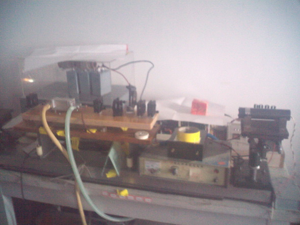
Since the bibliography of former experiments did not give any details about the optical system, so much work needs to be done to fill the blank. A He-Ne laser is employed as an indicator.YAG 1.06μm free running is applied. A large caliber(700μm) quartz fiber is used to transmit light to the operation platform of a stereomicroscope(45x). Specific collimation and focus system is designed to decrease the size of ablation spot.
|
|
outlet chamber |
|---|---|
| transverse mode selector | |
| Power supply of He-Ne Laser | |
| diaphragm | |
| holophote | |
| 1.06μm YAG | |
| YAG Power Supply and Pulse Frequency Control | |
| He-Ne |
1.06μm YAG
Fig 1, Laser Generator
Point to certain parts on the picture to see detail information
|
Focus lens |
|---|---|
| Fiber,700μm core diameter |
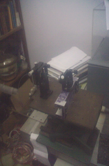
Fig 2. Fiber coupling
Point to certain parts on the picture to see detail information
|
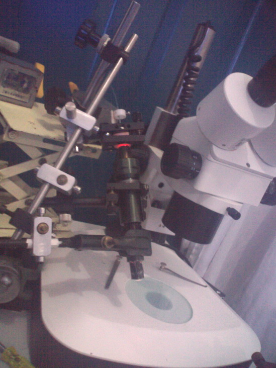
Stereoscope |
|---|---|
| Fiber Orientator | |
| Bean Expander (battery of lens consists of one positive len in the front and one negative len in the back) | |
| 3-demension adjust lever | |
| Focus len (f=10mm) | |
| Working Platform |
Fig 3. Focus System
Point to certain parts on the picture to see detail information
1.Ablation on photographic paper
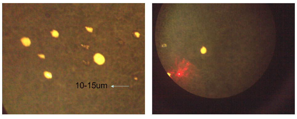
Fig 4. Ablation and Target spot
Left Pic - different sizes of ablation spots on black paper, the smallest is made at 270V; Right Pic - Red Spot or He-Ne target laser, spot size 20um
2.Ablation on leave cells

Fig 4. Ablation on Leave single cell
Left - Aiming; Right - Single cell destroyed after 3 pulses at 700V
Cell size: 20-30μm

Fig 5. Ablation a line on leave
Left - two layers of leave cells have been 'suddenly' destroyed at the last pulse of 10 consecutive pulses under 700V
Right- in the middle of the leave 500V pulse applied but only destroyed one layer of leave cells (other condition are the same of left)
The ditch width:≥100μm
3. Ablation standard tomato SAM
 |
 |
|---|
 |
 |
 |
|---|
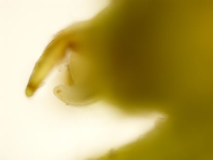
Fig 6. He-Ne targeting at SAM
Up Left - SAM Samples; Up Right- Original SAM; Down Left- Original SAM Marked with primordium P1 and P2
Down Mid- Turn off light and target SAM by using He-Ne laser(Red light); Down Right- Focus spot of red light
 |
 |
|---|
 |
 |
|---|
Fig 7 After Ablation
Up Left- Though Targeting in the center, but ablation mainly takes place at edge; Up Right- SAM continues to grow after ablation, so the ablation is not effective
Down Left- SAM under microscope from a side view; Down Right- SAM marked with its three primordia
Main problems and possible solutions
I would like to note here that I’m not biology major, nor an optics major of physics. So the information given above is a bit coarse and inaccurate, especially in its biological parts. I have had no cognition of Prof Huang Hai's hereditary theory before. Therefore, the experiment is somehow a challenge to me because much has to be learned and I’m the only one who mainly deals with the experiment on its physical part in Laser Biology Lab of USTC in Hefei. Xu Deyang from Shanghai Institute of Plant Physiology & Ecology of Chinese Academy of Sciences is responsible for biological issues such as cultivating and providing me tomato samples, cutting off SAM, continuing cultivating SAM after ablation and make slices of samples. Prof Li Yinmei offers me guidance on optical construction, and Prof Zhou Xiaonan takes part in assembling the YAG and He-Ne laser generator.
Because two teams work in different cities, the progress is somehow slow without adequate exchange. The experiment above is only a validating experiment. We get a conclusion from above, current application, and experiment done before, that tissues ablation is feasible. So our next step is to solve existing problems and improving experimental conditions.
The experiment requires the ablation spot to be 20μm in width and carve a line on tissues on SAM. However, the current experiment situation is far from reaching its required level. I have to illustrate here that the experiment is now at the very beginning – validating if it is possible to ablate tissues. And I try achieving the goal of accuracy at a minimal price. And doing the research from a much lower standard I can get a more basic understanding of the physical conditions and effects at the same time. And achievements can be made through analysis of conditions that do not meet the requirement. This is much better than blindly investing in advanced equipment to accomplish the experiment with unnecessary expenses.
By comparing laser ablation experiment made by people in the past with ours, the main difference is Q –switch and Er:YAG with 2.94μm wavelength. Other differences also contribute to different ablation effects. The following is my analysis.
After the validating experiment written above, we are assembling a Q-swtiched YAG laser and designing a flexible optical knife to focus the ablation spot. Further experiment result is expected ahead the end of this year.